Microtia and Anotia Ear Deformities: The Path from Diagnosis to Treatment
Seeing your newborn in your arms for the first time is the most beautiful sight you have ever seen, and it brings an unrivaled sense of delight. Your newborn’s coos and gurgles bring you unimaginable joy. Amidst all this happiness and relief, you may observe that your newborn’s ears are smaller than usual, possibly lacking some features, or one or both ears are absent entirely. Depending on the extent of ear deformity, the hospital pediatrician who checked your infant shortly after birth has talked to you about this or will soon.
Since you are quite likely at a loss to understand what this means for your child and what you can do to help your little one, this blog will help you understand what Microtia and Anotia are, what treatments are available, and where to turn for help.
The Definition of Microtia or Anotia Deformity
The Microtia ear deformity is a rare congenital birth defect whereby one or both of the external ears are either undeveloped or, in the case of Anotia, completely missing. It is often associated with hearing loss and/or facial abnormalities.
There are Four Grades of Microtia
- The ear is smaller than normal but still looks like an ear
- The ear is smaller and some ear features are missing
- The ear is severely underdeveloped, often with only a small remnant of ear present
- The ear is completely missing. This is called Anotia.
What Causes Microtia and Anotia Ear Deformities?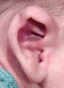
Although the precise origins of Microtia and Anotia are not well known, a number of factors have been found to be involved:
- Genetics
- In animal studies, Microtia and Anotia have been associated with mutations in genes such HOXA2, SIX, EYA, TBX1, IRF6, and CHUK1.
- Certain cases are inherited; if one child or biological family member has Anotia or Microtia, there is an approximate 5% chance that another child will also have the condition.
- Mutations: Genetic diseases like Treacher Collins syndrome, which is brought on by mutations in the TCOF1 gene1, can be linked to Microtia and Anotia.
- Environmental factors
- Maternal illness during pregnancy, particularly in the first trimester.
- Maternal diabetes before pregnancy.
- Exposure to teratogens such as retinoic acid, thalidomide, and mycophenolate mofetil1 .
- Use of isotretinoin (Accutane) during pregnancy.
- Abnormal vitamin A levels during pregnancy.
- Maternal diet low in carbohydrates and folic acid.
- Vascular disruption
- According to some researchers, microtia1 may result from localized ischemia and tissue necrosis (cell death) brought on by a breakdown in the blood supply while in the womb. In essence, the ear doesn’t get enough blood supply to develop or to continue to develop.
- Other elements
- Extreme mother or father age.
- Multiple births.
- Exposure to high altitudes.
- Native American or Hispanic heritage.
It is crucial to remember that microtia is a rare occurrence and is not caused by the mother’s actions or inactions during pregnancy.
How are Microtia and Anotia Deformities Diagnosed?
Although you may not understand what the deformity is called, you would immediately notice if your newborn has a partial or missing ear. Likely, it will be diagnosed (named) by the physician who first examines your baby in the newborn nursery.
The Treatment Option for Microtia and Anotia Ear Deformities
Microtia treatment typically involves a multidisciplinary approach, addressing both the physical appearance of the ear and any associated hearing loss. The main treatment options include the following.
Non-Surgical Options for Children with Microtia or Anotia
EarWell is not an option for infants with Microtia or Anotia, as it works only when an intact ear is present. However, prosthetic ears are a temporary or permanent option for those with Microtia or Anotia. These artificial ears are made of silicone that are attached to the head with either adhesive or magnets. While they do require regular maintenance and replacement as they are worn or the child grows, it does eliminate surgical reconstruction, which may be a risk for some children and adults.
Types of Surgical Reconstruction for Microtia and Anotia
Surgical reconstruction is the only permanent treatment of choice for both Microtia and Anotia, and is usually performed when the child is school-aged, though some procedures can be done as early as age 4. While not classified as cosmetic surgery, a surgical procedure for Microtia or Anotia does carry cosmetic advantages with it, since ear reconstruction not only helps your child hear better and speak more clearly, it can also help align their facial features and reduce self-consciousness, and diminish the inclination for other children to bully those who look different.
Autologous Reconstruction uses the patient’s own rib cartilage to create a new ear. The surgery is typically performed in stages, starting around age 7-10 when there’s enough rib cartilage available.
Synthetic Implants like MEDPOR are sometimes used to create the ear frame. This technique can sometimes be performed at a younger age than autologous reconstruction, since no rib cartilage is needed. An example of this option, in Dr. Sheryl Lewin’s case of a 3-year-old girl, can be viewed here.
The TPF Technique
The TPF (Temporoparietal Fascial Flap) Technique is used in microtia reconstruction to reduce scalp scars. Here’s a breakdown:
- How TPF Flaps Work:
- A flap of tissue and fascia (a strong connective tissue) is harvested from the scalp area, specifically the temporoparietal region.
- This flap is then used to cover the cartilage framework that has been created to form the new ear.
- Often, a thin skin graft is placed over the TPF flap to provide the final skin covering for the reconstructed ear.
- Why TPF Flaps are Used:
- Good Blood Supply: The TPF flap has a reliable blood supply, which is crucial for successful tissue survival.
- Hairless: The scalp area typically has minimal hair growth, minimizing the risk of hair growth on the reconstructed ear.
- Flexibility: The fascia provides a good foundation for the reconstructed ear, allowing for some flexibility and movement.
Note:
- This is a complex surgical procedure that requires specialized expertise.
- The specific techniques and approaches may vary depending on the individual case and the surgeon’s preference.
The Lewin Ear Implant
The Lewin Ear Implant is a significant advancement in microtia ear reconstruction. Here’s a summary:
- What it is: It’s a custom-made ear implant created from a single piece of porous polyethylene. This eliminates the risk of breakage that was associated with earlier two-piece implants.
- Key Features:
- 3D Scanning: High-definition 3D scanning is used to create a precise mirror image of the patient’s “good” ear or, in cases where both ears are affected, the ear of a sibling. This allows for highly individualized and natural-looking results.
- Single-Piece Design: The use of a single piece of material significantly reduces the risk of implant fracture, a major concern with older techniques.
- Improved Aesthetics: The Lewin Ear Implant is designed to achieve a more natural and aesthetically pleasing appearance compared to previous methods.
- Benefits:
- Reduced Risk of Complications: The single-piece design minimizes the risk of implant breakage, leading to fewer complications and the need for revision surgeries.
- Improved Outcomes: The 3D scanning technology allows for greater precision and customization, resulting in more natural-looking and symmetrical ears.
- Enhanced Patient Satisfaction: The improved aesthetics and reduced risk of complications can significantly enhance patient satisfaction with the surgical outcome.
3D Patient-Specific Porous Implant Ear Reconstruction is a cutting-edge technique that utilizes 3D printing technology to create customized ear implants for individuals with microtia. Here’s a breakdown:
- How it Works:
- 3D Scanning: A 3D scan of the patient’s “good” ear (or the ear of a sibling in cases of bilateral microtia) is obtained.
- Computer-Aided Design (CAD): The 3D scan data is used to create a mirror image of the normal ear using CAD software. This allows for the creation of a highly customized and anatomically accurate ear implant.
- 3D Printing: The 3D model is then used to 3D print the ear implant using biocompatible materials such as porous polyethylene. This allows for precise control over the shape, size, and contours of the implant.
- Surgical Implantation: The 3D-printed implant is surgically placed under the skin to form the framework of the reconstructed ear.
- Benefits:
- Highly Personalized: The 3D printing process allows for the creation of highly customized implants that closely match the shape and size of the patient’s normal ear, resulting in more natural-looking outcomes.
- Improved Aesthetics: The increased precision and customization can lead to significant improvements in the aesthetic appearance of the reconstructed ear.
- Reduced Complications: The use of biocompatible materials and precise 3D printing can minimize the risk of complications such as implant rejection or fracture.
- Key Considerations:
- This is a complex surgical procedure that requires specialized expertise in both 3D printing and microtia reconstruction.
- The long-term outcomes of this technique are still being studied.
Ear Canal Reconstruction: For patients with aural atresia (no ear canal), surgeons can create a new ear canal and eardrum to improve hearing.
Hearing Devices: For patients with hearing loss, options include:
- Bone-anchored hearing aids (BAHA)
- Vibrant sound bridge middle ear implants
Treatment Timing
The timing of treatment varies depending on the specific case:
- For unilateral microtia (affecting one ear), surgical reconstruction often starts around age 5-7
- For bilateral microtia (affecting both ears), treatment may begin earlier, around age 4, due to the increased risk of hearing and speech development issues.
Choosing a Treatment
The best treatment option depends on various factors, including:
- Severity of the microtia
- Presence of hearing loss
- Child’s age and overall health
- Family preferences
A team of specialists, including plastic surgeons, otolaryngologists, audiologists, and speech-language pathologists, typically work together to create a personalized treatment plan for each child
The Importance of Psychosocial Support for Children with Microtia/Anotia
Since these severe ear deformities can impact self-esteem and body image, psychological counseling or support groups may also be beneficial for patients and their families.
Go to our Provider Physician locator page, enter your location details and one or more specially trained providers should appear. If you have trouble using the system or can’t find a physician near enough, call us at 866-431-0480


